Scalable DICOM 3D-printed phantoms mimicking marine mammal bone and soft tissue
Abstract
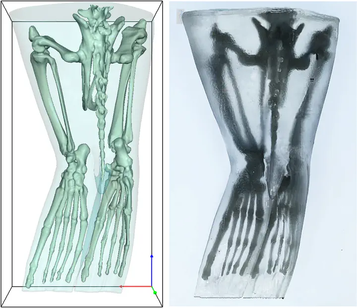
As charismatic sentinel species, California sea lions (Zalophus californianus) are commonly found in professional care settings such as zoos, aquariums, and rehabilitation facilities, in addition to their free-ranging coastal populations. These animals frequently strand due to illness, trauma, or environmental stressors, including toxic algal blooms such as domoic acid poisoning, underscoring the need for innovative tools and training methods to improve diagnostic care, monitoring, and veterinary intervention. This study presents a systematic approach for developing scalable, 3D-printable phantoms of a California sea lion pelvis using DICOM (Digital Imaging and Communications in Medicine) standard images from computed tomography (CT) scans to aid in veterinary blood collection training. The CT image data was processed using Simpleware ScanIP software to create detailed anatomical models, emphasizing the blood collection site at the caudal gluteal region and optimized for 3D printing. Through threshold-based segmentation of the DICOM data, several distinct anatomical layers were modeled separately, including a combined epidermal and dermal compliant skin shell, an adipose-rich blubber layer, a muscular layer derived from lower-density soft tissue regions, and a skeletal structure segmented from high-density bone data. This separation enabled each component to be fabricated independently using materials that closely matched their biological counterparts. Prior to fabrication, a material characterization study was conducted using dynamic mechanical analysis (DMA) to evaluate the compressive viscoelastic properties of multiple Humimic medical gelatin compositions (Gels 0 through 5), each with distinct mechanical profiles. The apparent elastic modulus of each gel under cyclic loading was calculated from stress–strain hysteresis data. Based on these results, individual gel types were selected to best match the mechanical properties of biological tissues, including blubber, skin, muscle, and bone. The quad-layered phantom was then fabricated using a combination of high-resolution stereolithography (SLA), fused deposition modeling (FDM), and gel casting techniques. This process resulted in the successful creation of 3D-printed anatomical phantoms that mimic both the mechanical and anatomical properties of the California sea lion pelvis. The methodology presented here provides a framework for creating engineered medical training models with anatomical fidelity and tunable material properties, offering a scalable alternative to traditional approaches in both veterinary and human health education, and the potential for personalized compatible implant design and biomimetic soft robotics.
Nazanin Minaian
Ph.D. in Mechanical Engineering | Research Faculty
My research interests include electroactive polymers, flow sensing, energy harvesting, and computer vision.